"We use cold plasma therapy and the CPT®patch to supplement traditional wound care for our difficult and poorly healing wounds."

Prof. Dr. med. Ole Goertz
Chief Physician for Plastic, Reconstructive & Aesthetic Surgery,
Martin Luther Hospital, Berlin
CPT® Plasma.Competence.Center.

for your treatment success

"We use cold plasma therapy and the CPT®patch to supplement traditional wound care for our difficult and poorly healing wounds."

Prof. Dr. med. Ole Goertz
Chief Physician for Plastic, Reconstructive & Aesthetic Surgery,
Martin Luther Hospital, Berlin
CPT® Plasma.Kompetenz.Zentrum.

Treatment with the CPT®patch

The application of the dressing is simple and safe

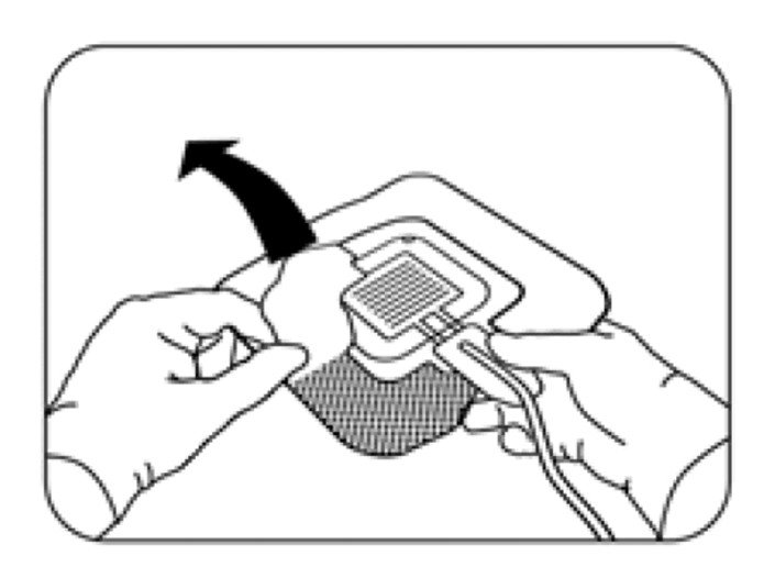
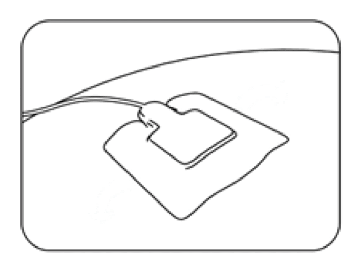
For treatment, apply a CPT®patch to the wound area and operate it by means of the CPT®cube.

For treatment, apply a CPT®patch to the wound area and operate it by means of the CPT®cube.

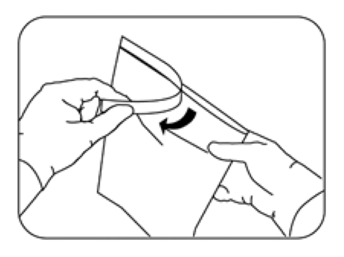
1. open sterile packaging

2. remove CPT®patch

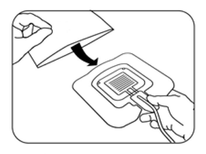
3. remove protective foil

3. apply CPT®patch

The treatment starts automatically after pressing the start/stop key on the CPT®cube. The treatment lasts 2 minutes.

The treatment can be repeated as often as necessary until the desired therapeutic success has been achieved. However, it should be noted that there should be at least 24 hours between each plasma treatment.

The treatment can and should be applied adjuvantly to other forms of therapy or recommendations of the corresponding guidelines at the discretion of the treating physician. Following plasma treatment, the wound is covered with commercially available wound dressings.

The effect

Clinical, dermatological examinations have revealed the following:

Application to the mode of action

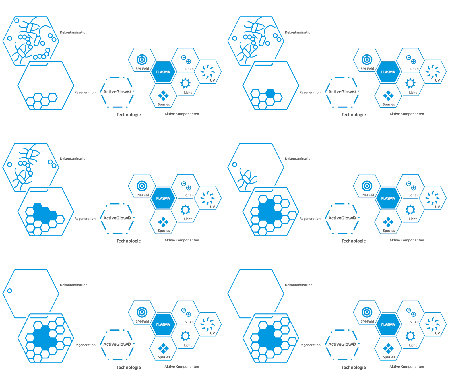
Application to the mode of action of disinfection using the example of COLDPLASMATECH

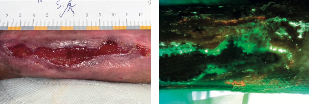
Initial situation: colonization with Pseudomonas aeruginosa, venous leg ulcer, 61-year-old patient
Initial situation - colonization with Pseudomonas aeruginosa
Result after treatment
with the CPT®patch
after treatment with the CPT®patch

The treatment and the systems

How does a cold plasma treatment work? 

Cold plasma is applied to the affected area of the skin in a flat or punctual manner. The treatment is performed 2-3 times a week. Cold plasma treatment is a gentle, easy-to-perform therapy. It is performed without pain and with no or light skin contact. Two forms of cold plasma treatment have been used so far:

Hand guided systems

The hand-applied cold plasma devices with variously sized attachments (for single use) treat areas ranging from pinpoints to several square centimeters. There are pen-shaped to stamp-like solutions. What they all have in common is the influence of the practitioner on the treatment. Care must be taken to avoid over- or under-treatment. The applicators are sometimes grated through the wound in contact with the wound.

Automatic systems

The treatment starts automatically after pressing the start/stop key. The treatment runs at the optimum effect and ends automatically after approx. 2 minutes. Due to the automatic treatment, over- or under-treatment is excluded. The adhesive edge on the applicator has atraumatic properties and prevents direct contact with the wound. The treatment is also suitable for large-area wounds.

The type and duration of plasma therapy depends on the particular wound conditions. In severely necrotic or inflamed wounds, plasma therapy should initially be used to accompany wound debridement and possible local or systemic drug therapy.

The intensity of the plasma treatment is controlled by the duration of the session and is repeated two to three times a week.

After the third treatment with theCPT®patch you could see - the wound is starting to heal really great. I can't see the cold plasma, but it smells like fresh air and the treatment is the first one that really helps me. 


Patient Ruth Wollgast after successful treatment with the CPT®patch

Why does plasma therapy work for wounds?

Cold plasma destroys germs, improves oxygen supply in the skin, stimulates cell growth and promotes wound healing.

The most important mechanism of action of plasma in wound treatment is probably its strong antimicrobial and anti-inflammatory (anti-inflammatory) effect. Medical cold plasma is only 37 °C warm. Nevertheless, it kills viruses, bacteria, fungi or spores within a few minutes.

In addition, the cold plasma stimulates cell division (cell proliferation) and cell migration. These processes increase collagen synthesis and support physiological wound healing. Even in the case of long-standing chronic wounds, cold plasma treatment achieves good treatment results. Possible side effects or pain during treatment have not occurred so far.

We use cold plasma therapy and the CPT®patch for our difficult and poorly healing wounds as a supplement to classic wound care.


Prof. Dr. Ole Goertz, Chief Physician Martin Luther Hospital, Berlin

Tips and tricks

Tips and tricks for treatment with the CPT®patch

The CPT®Patch - structure & application

Play video

Treatment of the lower leg with the CPT®Patch

Play video

Treatment of the toe with the CPT®Patch

Play video

Treatment of the heel with the CPT®Patch

Play video

Treatment of difficult areas, treatment of Bisgaard's backdrop with the CPT®Patch

Play video

The treatment and the systems

How does a cold plasma treatment work? 

Cold plasma is applied to the affected area of the skin in a flat or punctual manner. The treatment is performed 2-3 times a week. Cold plasma treatment is a gentle, easy-to-perform therapy. It is performed without pain and with no or light skin contact. Two forms of cold plasma treatment have been used so far:

Hand guided systems

The hand-applied cold plasma devices with variously sized attachments (for single use) treat areas ranging from pinpoints to several square centimeters. There are pen-shaped to stamp-like solutions. What they all have in common is the influence of the practitioner on the treatment. Care must be taken to avoid over- or under-treatment. The applicators are sometimes grated through the wound in contact with the wound.

Automatic systems

The treatment starts automatically after pressing the start/stop key. The treatment runs at the optimum effect and ends automatically after approx. 2 minutes. Due to the automatic treatment, over- or under-treatment is excluded. The adhesive edge on the applicator has atraumatic properties and prevents direct contact with the wound. The treatment is also suitable for large-area wounds.

The type and duration of plasma therapy depends on the particular wound conditions. In severely necrotic or inflamed wounds, plasma therapy should initially be used to accompany wound debridement and possible local or systemic drug therapy.

The intensity of the plasma treatment is controlled by the duration of the session and is repeated two to three times a week.

After the third treatment with theCPT®patch you could see - the wound is starting to heal really great. I can't see the cold plasma, but it smells like fresh air and the treatment is the first one that really helps me. 


Patient Ruth Wollgast after successful treatment with the CPT®patch

Why does plasma therapy work for wounds?

Cold plasma destroys germs, improves oxygen supply in the skin, stimulates cell growth and promotes wound healing.

The most important mechanism of action of plasma in wound treatment is probably its strong antimicrobial and anti-inflammatory (anti-inflammatory) effect. Medical cold plasma is only 37 °C warm. Nevertheless, it kills viruses, bacteria, fungi or spores within a few minutes.

In addition, the cold plasma stimulates cell division (cell proliferation) and cell migration. These processes increase collagen synthesis and support physiological wound healing. Even in the case of long-standing chronic wounds, cold plasma treatment achieves good treatment results. Possible side effects or pain during treatment have not occurred so far.

We use cold plasma therapy and the CPT®patch for our difficult and poorly healing wounds as a supplement to classic wound care.


Prof. Dr. Ole Goertz, Chief Physician Martin Luther Hospital, Berlin

The active components explained

Active components (excerpt from position paper on risk potential and application perspectives of cold atmospheric pressure plasma in medicine, NZPM)

According to the current international state of research, the main active components of cold atmospheric pressure plasmas are reactive nitrogen and oxygen species (RNS, ROS), UV radiation and electric fields.

According to the current international state of research, the main active components of cold atmospheric pressure plasmas are reactive nitrogen and oxygen species (RNS, ROS), UV radiation and electric fields.

Reactive nitrogen and oxygen species (RNS, ROS) are formed locally and briefly by coupling electrical energy into gases that are not biologically active per se (argon, helium, nitrogen, oxygen, air and mixtures thereof) and subsequent interaction with adjacent media (atmospheric air, liquids, surfaces). In principle, the same reactive species are sometimes produced in the human body as part of normal metabolism and in some cases have important functions in controlling and mediating physiological and pathological processes. Briefly elevated doses of these RNS and ROS can be effectively detoxified by the body's own systems.

UV radiation is used medically in phototherapy and photochemotherapy, among other things. In this context, as well as under the aspect of general personal and occupational safety, also outside the medical environment, limit values have been defined for UV exposure, which are significantly undercut in connection with the use of cold atmospheric pressure plasma. 

Two important findings of basic plasma medicine research in recent years are:

1. biological plasma effects on cells and in tissues are mediated via changes in the fluid cell environment.

2. for biological effects induced by plasma action, oxidizing species, so-called reactive oxygen and nitrogen species (ROS, RNS), introduced into the liquid or formed in the liquid play a dominant role.

The same reactive species (ROS, RNS) are also produced in the human body as part of normal metabolism and in some cases have important functions in controlling and mediating physiological and pathological processes. The most important ROS and RNS are hydroxyl radical (OH-), hydrogen peroxide (H2O2), superoxide or hyperoxide (O2--), nitric oxide (NO-), nitrogen dioxide (NO2-) and peroxynitrite (ONOO-). They play an important role in wound healing processes, for example. This finding provides an essential scientific basis for the concept of plasma-assisted wound healing, in which, in addition to the well-known antibacterial/disinfectant plasma effect, stimulation of tissue regeneration is also to be achieved by plasma action. One mechanism of plasma action is thus based on the support of endogenous functions which - for example in the case of non-healing chronic wounds - cannot be sufficiently effective due to disease-related disorders. Furthermore, it is known that a certain basic concentration of RNS and ROS is always present in human cells. Due to the physiological occurrence of these species, short-term elevated concentrations can be effectively detoxified by endogenous systems [16-24]. Transcriptome analyses of in vitro plasma-treated human cells have shown that genes associated with the cellular stress response are increasingly upregulated and antioxidant active enzymes are produced as a result of plasma treatment [25]. 

Since plasma treatments are local and time-limited, under normal conditions the risk of side effects associated with entry of these ROS and RNS into the tissue is expected to be exceptionally low. 

3. ultraviolet radiation (UV radiation)

UV-B radiation in particular is used in dermatology as part of phototherapy. According to the recommendations of the German Dermatological Society (DDG) on phototherapy and photochemotherapy, initial doses of between 20 and 60 mJ/cm2 are recommended for broad-spectrum UV-B application (280-320 nm) depending on the skin type, and doses of between 200 and 600 mJ/cm/WTA are recommended for narrow-spectrum UV-B treatment (311 nm).

A comparison with solar radiation shows that the UV intensity emitted by the cold atmospheric pressure plasma sources used in clinical testing or approved as medical devices to date is far below that of sunlight.

For the plasma sources currently certified as medical devices, it has been shown that under the recommended conditions of use (working distance, treatment time), the maximum permissible daily UV dose is significantly undercut.


4. electric fields

Electric fields can first be divided into direct and alternating fields. Furthermore, the pulsation of such signals as well as a mono- or bi-phase modulation allows a high parametric diversity. Technical frequencies are in the range of a few Hz up to the GHz range. Devices that work exclusively with electric fields have been established for many years for use in and on the human body and can induce an electric current flow in biological tissue as a result of the electric fields. Regardless of the device, the use of electrical signals for electrostimulation offers a variety of proven applications in medical care. For example, cell movement of immune cells (macrophages and granulocytes) and migration of skin cells (keratinocytes) and corneal epithelium can be specifically influenced in response to an electric field, proliferation behavior of connective tissue cells (fibroblasts) is stimulated, and for new vessel formation (angiogenesis) and nerve growth, the electric field is also important. Finally, in vivo studies using electric fields have shown an antibacterial effect on both gram-negative and gram-positive bacteria.

In recent years, research in the field of bioelectricity has demonstrated significant links between endogenous electric fields and the wound healing process. A meta-analysis by Gardner et al. was performed using data sets from 15 clinical trials with the goal of quantifying the effect of electrical stimulation (ES) on chronic wound healing. Treatments with ES achieved an average wound reduction of 22.2% per week, compared with only 9.1% in control groups. In the published sub-results of Cochrane Review #077, healing success (wound closure) using electrical stimulation was compared to placebo control. This question was investigated in 13 of the 20 studies. The analysis showed that twice as many (OR=2.12; 95% CI: 1.55 - 2.90) wounds were healed by treatment with electrostimulation (verum) compared to controls.

Placeholder

  1. Sagawa et al, Argon plasma coagulation for successful treatment of early gastric cancer with intramucosal invasion. Gut 52 (2003) 334-339
  2. J. Vargo, Clinical applications of the argon plasma coagulator. Gastrointestinal Endoscopy 59 (2004) 81-88
  3. Raiser, M. Zenker. Argon plasma coagulation for open surgical and endoscopic applications: state of the art. J. Phys. D: Appl. Phys. 39 (2006) 3520-3523
  4. Manner et al, Safety and efficacy of a new high power argon plasma coagulation system (hp-APC) in lesions of the upper gastrointestinal tract. Digestive and Liver Disease 38 (2006) 471-478.
  5. Manner et al, Second-generation argon plasma coagulation: Two-center experience with 600 patients. J. Gastroenterol. Hepatol. 23 (2008) 872-878.
  6. Zenker, Argon plasma coagulation. GMS Hospital Hyg. Interdisct. 3 (2008)Doc15
  7. Keller et al, Electrical and spectroscopic characterization of a surgical argon plasma discharge. J. Phys. D: Appl. Phys. 46 (2013) 025402
  8. Keller, Characterization of argon plasma coagulation (APC) for thermal treatment of biological tissue in endoscopy and surgery. Dissertation for the degree of Doctor of Engineering at the Faculty of Electrical Engineering and Information Technology, Ruhr University Bochum 2012.
  9. Ianelli et al, Use of PlasmaJet System in patients undergoing abdominal lipectomy following massive weight loss resulting from bariatric surgery: early experience. Obes. Surg. 16 (2006) 1504-1507
  10. Nezhat et al, Use of neutral argon plasma in the laparoscopic treatment of endometriosis. J. Soc. Laparoendsocop. Surg. 13 (2009) 479-483
  11. K. Madhuri, et al, First clinical experience of argon neutral plasma energy in gynaecological surgery in the UK. Gynaecol. Surg. 7 (2010) 423-425.
  12. A. Bogle et al, Evaluation of plasma skin regeneration technology in low-energy full-facial rejuvenation. Arch. Dermatol. 143 (2007) 168-174.
  13. Kilmer et al, A pilot study on the use of a plasma skin regeneration device (Portrait PSR3) in full facial rejuvenation procedures. Lasers Med. Sci. 22 (2007) 101-109
  14. D. Holcomb et al, Nitrogen plasma skin regeneration and aesthetic facial surgery. Arch. Facial Plast. Surg. 11 (2009) 184-193
  15. Wade Forster et al, Advances in plasma skin regeneration (Review article). J. Cosmetic Dermatol. 7 (2008) 169-179.
  16. Dröge, Free radicals in the physiological control of cell function. Physiol. Rev. 82 (2002) 47-95
  17. C. Fang, Antimicrobial reactive oxygen and nitrogen species: concepts and controversies. Nature Rev. Microbiol. 2 (2004) 820-832
  18. G. Rhee, H2O2, a necessary evil for cell signaling. Science 312 (2006) 1882-1883
  19. O. Lundberg et al, The nitrate-nitrite-nitric oxide pathway in physiology and therapeutics. Nature Rev. Drug Discov. 7 (2008) 156-168
  20. K. Sen and S. Roy, Redox signals in wound healing (invited review). Biochim. Biophys. Acta 1780 (2008) 1348-1361
  21. K. Sen, Wound healing essentials: let there be oxygen (perspective article). Wound Rep. Regen. 17 (2009) 1-18
  22. Leonarduzzi et al, Targeting tissue oxidative damage by means of cell signaling modulators: the antioxidant concept revisited. Pharmacology & Therapeutics 128 (2010) 336-374
  23. B. Graves, The emerging role of reactive oxygen and nitrogen species in redox biology and some implications for plasma applications to medicine and biology (topical review). J. Phys. D: Appl. Phys. 45 (2012) 163001
  24. Von Woedtke et al, Plasmas for medicine. Phys. Rep. 530 (2013) 291-320
  25. Schmidt et al. Non-thermal plasma treatment is associated with changes in transcriptome of human epithelial skin cells. Free Radical Res. 47 (2013) 577-592
  26. Kramer, O. Assadian (Eds.), Praxis der Sterilisation, Desinfektion, Antiseptik und Konservierung, Thieme, Stuttgart 2008, pp. 719 ff.
  27. Fachverband für Strahlenschutz e.V., Guideline "Ultraviolet radiation from artificial sources" (FS-2013-157-AKNIR, as of 28.3.2013); http://osiris22.pi-consult.de/userdata/l_20/p_105/library/data/fs-05-131-aknir_uv_strahlung.pdf
  28. Directive 2006/25/EC of the European Parliament and of the Council of 5 April 2006 on the minimum health and safety requirements regarding the exposure of workers to the risks arising from physical agents (artificial optical radiation); http://eur-lex.europa.eu/LexUriServ/LexUriServ.do?uri=OJ:L:2006:114:0038:0059:de:PDF
  29. ICNIRP Statement. Guidelines on UV radiation exposure limits. Health Physics 87 (2004) 171
  30. Association of the Scientific Medical Societies (AWMF), AWMF Guideline Register No. 013/029, Recommendations of the German Dermatological Society (DDG) on UV phototherapy and photochemotherapy, as of 12/2009; http://www.awmf.org/uploads/tx_szleitlinien/013-029.pdf
  31. Lademann et al, Risk assessment of the application of a plasma jet in dermatology. J. Biomed. Opt. 14 (2009) 054025
  32. Heinlin et al, Plasma applications in medicine with a special focus on dermatology. J. Eur. Acad. Venereol. 25 (2011) 1-11
  33. Isbary et al, Successful and safe use of 2 min cold atmospheric argon plasma in chronic wounds: results of a randomized controlled trial Br. J. Dermatol. 167 (2012) 404-410.
  34. Rajasekaran et al, Characterization of Dielectric Barrier Discharge (DBD) on Mouse and Histological Evaluation of the Plasma-Treated Tissue. Plasma Process. Polym. 8 (2011) 246-255.
  35. Bussiahn et al, Plasma sources for biomedical applications. Hyg. Med. 38 (2013) 212-216
  36. R. Cho et al, Integrin-Dependent Human Macrophage Migration Induced by Oscillatory Electrical Stimulation Ann. Biomed. Eng. 28 (2000) 234-243
  37. Goldman, S. Pollack, Electric fields and proliferation in a chronic wound model. Bioelectromagnetics 17 (1996) 450-457.
  38. Song et al, Nerve regeneration and wound healing are stimulated and directed by an endogenous electrical field in vivo. J. Cell Sci. 117 (2004) 4681-4690
  39. E. Pullar et al, β4 Integrin and Epidermal Growth Factor Coordinately Regulate Electric Field-mediated Directional Migration via Rac1. Mol. Biol. Cell 17 (2006) 4925-4935.
  40. Daeschlein et al, Antibacterial activity of positive and negative polarity low-voltage pulsed current (LVPC) on six typical Gram-positive and Gram-negative bacterial pathogens of chronic wounds. Wound Repair Regen. 15 (2007) 399-403
  41. C. Recio et al, High-voltage Electrical Stimulation for the Management of Stage III and IV Pressure Ulcers among Adults with Spinal Cord Injury: Demonstration of its Utility for Recalcitrant Wounds below the Level of Injury, J. Spinal Cord. Med. 35 (2012) 58-63
  42. E. Houghton et al, Electrical Stimulation Therapy increases rate of healing of Pressure Ulcers in Community-Dwelling People with Spinal Cord Injury, Arch. Phys. Med. Rehabil. 91 (2010) 669-678.
  43. Adunsky et al. Decubitus Direct Current Treatment of Pressure Ulcers: Results of a Randomized, Double-Blind, Placebo-controlled Study, Arch. Gerontol. Geriatr. 41 (2005) 261-269
  44. E. Gardner et al, Effect of electrical stimulation on chronic wound healing: a meta-analysis. Wound Repair Regen. 7 (1999) 495-503
  45. Koel and F. Oosterveld. Review. Electrotherapy for stimulation of wound healing. EWMA Journal 9 (2009) 57
  46. DIN EN 60601-1. Medical electrical equipment - Part 1: General requirements for safety including essential performance. Issue date 2013-12
  47. ICNIRP Statement. Guidelines for limiting exposure to time-varying electric, magnetic, and electromagnetic fields (up to 300 GHz). Health Physics 97 (2009) 257-258
  48. Isbary et al, Ex vivo human skin experiments for the evaluation of safety of new cold atmospheric plasma devices. Clin. Plasma Med. 1 (2013) 36-44
  49. Kalghatgi et al, Effects of Non-Thermal Plasma on Mammalian Cells. PLoS ONE 6 (2011) e16270
  50. OECD 476/1997: OECD Guidelines for the testing of chemicals. In Vitro Mammalian Cell Gene Mutation Test; http://www.oecd.org/chemicalsafety/risk-assessment/1948426.pdf
  51. Boxhammer et al, Investigation of the mutagenic potential of cold atmospheric plasma at bactericidal dosages. Mutation Res. 753 (2013) 23-28
  52. Arndt et al. Cold Atmospheric Plasma (CAP) Changes Gene Expression of Key Molecules of the Wound Healing Machinery and Improves Wound Healing In Vitro and In Vivo. PloS One 8 (2013) e79325
  53. Isbary et al, A first prospective randomized controlled trial to decrease bacterial load using cold atmospheric argon plasma on chronic wounds in patients. Brit. J. Dermatol. 163 (2010) 78-82
  54. Isbary et al, Cold atmospheric plasma: A successful treatment of lesions in Hailey-Hailey disease. Arch. Dermatol. 147 (2011) 388-390
  55. Isbary et al, Non-thermal plasma-More than five years of clinical experience. Clin. Plasma Med. 1 (2013) 19-13
  56. Isbary et al, Cold atmospheric argon plasma treatment may accelerate wound healing in chronic wounds: results of a retrospective in vivo randomized controlled study. Clin. Plasma Med. 2 (2013) 25-30
  57. Isbary et al, Cold atmospheric plasma for local infection control and subsequent pain reduction in a patient with chronic post-operative ear infection. New Microb. New Inf. 3 (2013) 41-43
  58. Heinlin et al, A randomized two-sided placebo-controlled study on the efficacy and safety of atmospheric non-thermal argon plasma for pruritus. J. Eur. Acad. Dermatol. Venereol. 27 (2013) 324-331
  59. Heinlin et al, Randomized placebo-controlled human pilot study of cold atmospheric argon plasma on skin graft donor sites. Wound Rep. Regen. 21 (2013) 800-807
  60. -R. Metelmann et al, Experimental Recovery of CO2-Laser Skin Lesions by Plasma Stimulation. Am. J. Cosmetic Surg. 29 (2012) 52-56.
  61. -R. Metelmann et al, Scar formation of laser skin lesions after cold atmospheric pressure plasma (CAP) treatment: A clinical long term observation Clin. Plasma Med. 1 (2013) 30-35
  62. Emmert et al, Atmospheric pressure plasma in dermatology: Ulcer treatment and much more. Clin. Plasma Med. 1 (2013) 24-29
  63. Tiede et al, Plasma applications: a dermatological view. Contrib. Plasma Phys. 54 (2014) 118-130
  64. Emmert et al, Treatment of Chronic Venous Leg Ulcers with a Hand-Held DBD Plasma Generator. Plasma Medicine (2014) DOI: 10.1615/PlasmaMed.2013005914
  65. Lademann et al, Risk assessment of the application of tissue-tolerable plasma on human skin. Clin. Plasma Med. 1 (2013) 5-10
  66. Kramer et al, Suitability of tissue tolerable plasmas (TTP) for the management of chronic wounds. Clin. Plasma Med. 1 (2013) 11-18
  67. Clinical Plasma Medicine Core Group. Clinical plasma medicine - position and perspectives in2012. Paper of consent, result of the workshop "Clinical Concepts in Plasma Medicine", Greifswald April 28th, 2012. Plasma Med. 1 (2013) 3-4.
  68. von Woedtke et al, Clinical Plasma Medicine: State and perspectives of in vivo application of cold atmospheric plasma. Contrib. Plasma Phys. 54 (2014) 104-117.
  69. Schlegel et al, Plasma in cancer treatment. Clin. Plasma Med. vol. 1 no. 2 (2013) 2-7
  70. Rupf et al. Removing biofilms from microstructured titanium ex vivo: a novel approach using atmospheric plasma technology. PLoS One. 10 (2011) e25893
  71. A.N. Idlibi et al. Destruction of oral biofilms formed in situ on machined titanium (Ti) surfaces by cold atmospheric plasma. Biofouling 29 (2013) 369-79]

Side effects

Side effects/tolerance of cold plasma

At the current state of clinical research, no clinically relevant side effects are known.

At the current state of clinical research, no clinically relevant side effects are known.

Study situation on cold plasma

An extensive body of studies shows the effectiveness

The antimicrobial efficacy was confirmed by further studies. Chronic wounds of 34 patients were treated with cold plasma or in combination with a wound antiseptic. The combination therapy showed the best efficacy [1].

In a monocenter, randomized-controlled clinical trial, seven patients each with at least 12-week-old chronic ulcers underwent plasma treatment or no treatment in addition to normal wound care. The plasma source was a dielectric-restrained discharge powered by air. Wound healing occurred similarly to standard therapy, while wound areas colonized with bacteria decreased by a mean of 88% in wounds treated with Palsma [2].

In another study with five subjects, two artificial wounds were created on each arm using negative pressure and the healing process was observed. The four wounds received either no treatment (A) a treatment with cold plasma (B), Octenisept© (C) or a plasma treatment followed by a treatment with Octenisept© (D). Normalized to initial area, plasma-treated wounds (B) healed fastest at all measured time points, while untreated wounds (A) healed slowest [3].

Sixteen patients included in the study (ten women and six men) with chronic leg ulcers were each treated with cold plasma three times a week over a period of two weeks. In addition to measuring antimicrobial activity, the aim of the study was to investigate the effect on wound healing. 

The parameters compared were, in addition to the number of bacterial colonies per square centimeter, the size of the wound surface and the change in wound volume. The authors concluded that the immediate antimicrobial effect of the two treatment methods was largely comparable. Plasma therapy was very well tolerated by patients and, according to the authors, is unlikely to cause allergies due to its physical principle of action [4].

In a more extensive study with 70 patients, a tendency towards improved healing was determined on chronic ulcers treated with cold plasma - compared to untreated wounds [5]. In addition, another randomized-controlled trial with 40 patients showed significantly improved healing after plasma treatment of acute wounds after skin grafts [6]. A summary of the current status of plasma application in animal experiments, in vivo, and clinical studies and case reports is also the subject of a recent review [7].

A risk assessment with reference to plasma species (temperature, UV radiation and free radicals) did not reveal any increased risks for humans [8].

Based on the state of clinical research, plasma applications in dermatology as well as plastic and aesthetic surgery currently have the highest prospects of success. The use of antimicrobial plasma effects, plasma-assisted stimulation of tissue regeneration and inflammation-modulating plasma effects are the focus of therapeutic indications.

Placeholder

[1] M. Klebes, C. Ulrich, F. Kluschke, A. Patzelt, S. Vandersee, H. Richter, A. Bob, J. Hutten, J.T. Krediet, A. Kramer, Combined antibacterial effects of tissue-tolerable plasma and a modern conventional liquid antiseptic on chronic wound treatment, Journal of biophotonics, 8 (2015) 382-391.

[2] F. Brehmer, H. Haenssle, G. Daeschlein, R. Ahmed, S. Pfeiffer, A. Görlitz, D. Simon, M. Schön, D. Wandke, S. Emmert, Alleviation of chronic venous leg ulcers with a hand-held dielectric barrier discharge plasma generator (PlasmaDerm® VU-2010): results of a monocentric, two-armed, open, prospective, randomized and controlled trial (NCT01415622), Journal of the European Academy of Dermatology and Venereology, 29 (2015) 148-155.

[3] S. Vandersee, H. Richter, J. Lademann, M. Beyer, A. Kramer, F. Knorr, B. Lange-Asschenfeldt, Laser scanning microscopy as a means to assess the augmentation of tissue repair by exposure of wounds to tissue tolerable plasma, Laser Physics Letters, 11 (2014) 115701.

[4] C. Ulrich, F. Kluschke, A. Patzelt, S. Vandersee, V. Czaika, H. Richter, A. Bob, J. von Hutten, C. Painsi, R. Hügel, Clinical use of cold atmospheric pressure argon plasma in chronic leg ulcers: A pilot study, Journal of wound care, 24 (2015).

[5] G. Isbary, W. Stolz, T. Shimizu, R. Monetti, W. Bunk, H.U. Schmidt, G.E. Morfill, T.G. Klämpfl, B. Steffes, H.M. Thomas, J. Heinlin, S. Karrer, M. Landthaler, J.L. Zimmermann, Cold atmospheric argon plasma treatment may accelerate wound healing in chronic wounds: results of an open retrospective randomized controlled study in vivo, Clinical Plasma Medicine, 1 (2013) 25-30.

[6] J. Heinlin, J.L. Zimmermann, F. Zeman, W. Bunk, G. Isbary, M. Landthaler, T. Maisch, R. Monetti, G. Morfill, T. Shimizu, J. Steinbauer, W. Stolz, S. Karrer, Randomized placebo-controlled human pilot study of cold atmospheric argon plasma on skin graft donor sites, Wound Repair Regen, 21 (2013) 800-807.

[7] T. Von Woedtke, H.R. Metelmann, K.D. Weltmann, Clinical plasma medicine: state and perspectives of in vivo application of cold atmospheric plasma, Contributions to Plasma Physics, 54 (2014) 104-117.

[8] J. Lademann, H. Richter, A. Alborova, D. Humme, A. Patzelt, A. Kramer, K.-D. Weltmann, B. Hartmann, C. Ottomann, J.W. Fluhr, Risk assessment of the application of a plasma jet in dermatology, Journal of biomedical optics, 14 (2009) 054025-054025-054026.
G-BA paves the way for reimbursement and decides on trial study

The evaluation procedure at the Federal Joint Committee (G-BA) for the treatment of chronic wounds with cold plasma has been ongoing for almost two years. Now (February 2023), the G-BA has decided in its plenary session to conduct a trial study for cold plasma therapy in chronic wounds for reimbursement. This clears the last hurdle on the way to reimbursement of wound treatment with cold plasma, from which millions of people in Germany alone could benefit.

In order for a healthcare service to be reimbursable by statutory health insurers in Germany, the Federal Joint Committee must first conduct an evaluation procedure and assess the medical and economic benefits of the therapy. For almost two years, manufacturers and leading wound experts have been driving the process for reimbursement of cold plasma therapy at the GBA. The decision to conduct the trial represents the most important milestone.

All participants in the plenary session at the G-BA agreed that cold plasma can positively influence the healing process of chronic wounds and will improve the quality of life of many people, as cold plasma therapy is superior to standard therapy. In addition to inpatient treatment, the focus is on the transsectoral treatment of chronic wounds in the office-based and outpatient setting.

"On July 15, 2021, the G-BA gave a positive decision on the application for a trial of cold plasma treatment for chronic wounds. According to this, the method demonstrates the potential of a necessary treatment alternative compared to standard wound treatment according to guideline recommendations. This assessment is largely based on the findings on the plausible and established surrogate endpoint change in wound size, which were taken from the studies supporting the application."

Citation §2.1 Appendix 2 TG Erp RL_ER 21 00 Cold Plasma

Placeholder

The Federal Joint Committee (G-BA) is the highest decision-making body of the joint self-government in the German healthcare system.

In the form of guidelines, it determines which medical services the approximately 73 million insured persons can claim. In addition, the G-BA decides on quality assurance measures for practices and hospitals.
Body of the joint self-government
In Germany, the legislator makes the fundamental decisions on the entitlement to benefits of the statutory health insurants.
The legislator has entrusted the G-BA as the highest body of the joint self-government with the task of specifying the so-called benefits catalog of the health insurance funds according to the generally recognized state of medical knowledge. The Federal Ministry of Health is responsible for legal supervision.
The G-BA is formed by the four major self-governing organizations in the health care system:

  • National Association of Statutory Health Insurance Physicians
    (KBV)
  • National Association of Statutory Health Insurance Dentists (KZBV)
  • German Hospital Federation (DKG)
  • Spitzenverband Bund (GKV-Spitzenverband)
    of health insurance funds

In accordance with the provisions of the Fifth Book of the Social Code, organizations that represent the interests of patients and self-help groups for chronically ill and disabled people at the federal level have the right to participate in consultations and submit proposals, but no voting rights.
The main task of the G-BA is to define the content of health care in more detail in guidelines and to decide which services are paid for by the SHI.
In this way, the G-BA fulfills its legal mandate to ensure that all patients with statutory health insurance can receive good care and benefit from medical progress. At the same time, its guidelines serve to ensure that care is provided in a quality-assured and economical manner.

Our goal: the healing of chronic wounds.

Our solution: therapy with cold plasma.

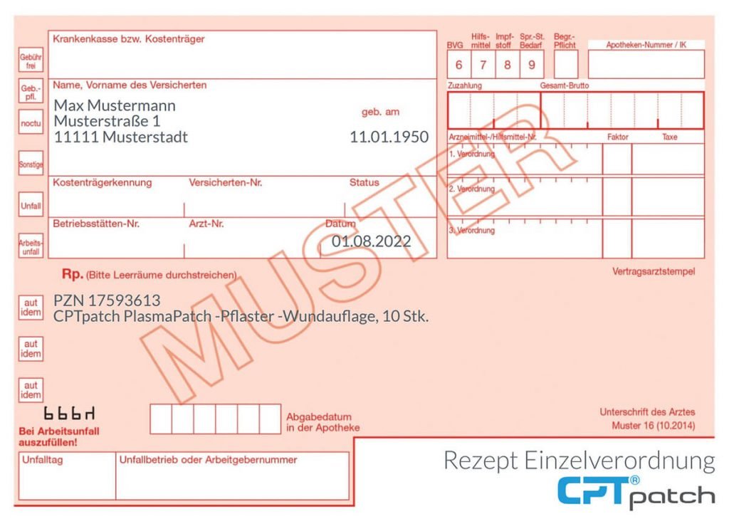
Reimbursement

If you have any questions regarding reimbursement, please contact us.

You can find information on data protection here

If you have any questions regarding reimbursement, please contact us.
You can find information on data protection here